41 bacterial cell picture with labels
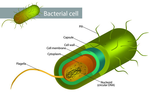
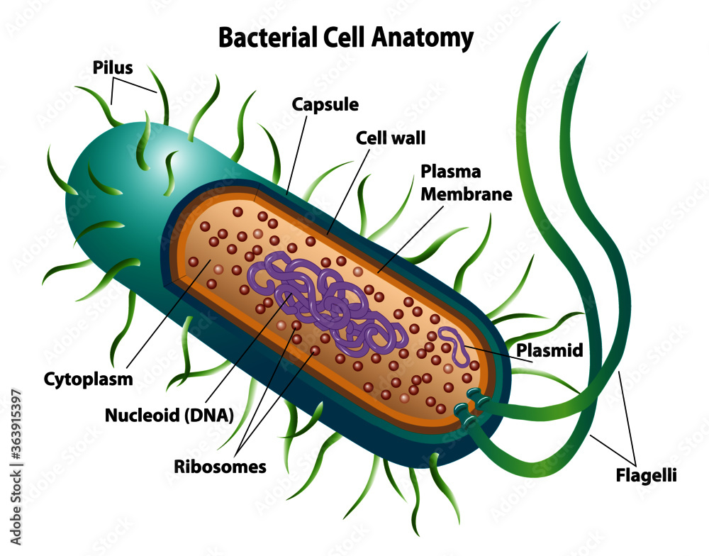
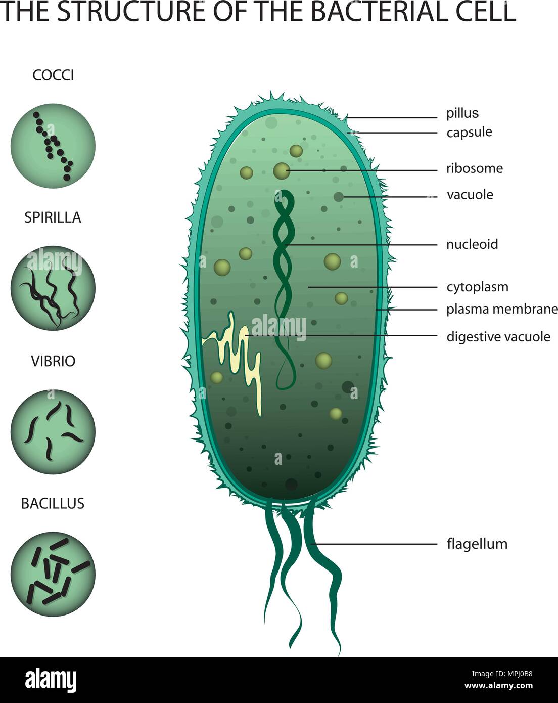
Animal Cell - Free printable to label + Color -kidCourses.com Can you label and color these important parts of the animal cell?. NUCLEUS control center for cell (cell growth, cell metabolism, cell reproduction). NUCLEOLUS synthesizes rRNA. RIBOSOMES the site of protein building, this is where translation takes place (mRNA in language of nucleic acids is translated into the language of amino acids). RER (Rough Endoplasmic Reticulum) synthesizes proteins ... Interactive Bacteria Cell Model - CELLS alive In the space are enzymes and other proteins that help digest and move nutrients into the cell. Cell Wall: Composed of peptidoglycan (polysaccharides + protein), the cell wall maintains the overall shape of a bacterial cell. The three primary shapes in bacteria are coccus (spherical), bacillus (rod-shaped) and spirillum (spiral).
Consumer Updates | FDA - U.S. Food and Drug Administration Jul 28, 2022 · The site is secure. The https:// ensures that you are connecting to the official website and that any information you provide is encrypted and transmitted securely.
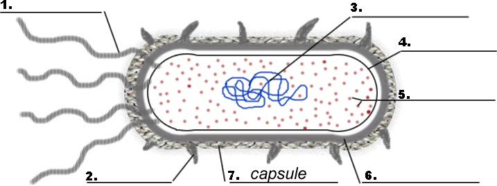
Bacterial cell picture with labels
Bacterial cells - Cell structure - Edexcel - GCSE Combined Science ... Feature Eukaryotic cell (plant and animal cell) Prokaryotic cell (bacterial cell) Size: Most are 5 μm - 100 μm: Most are 0.2 μm - 2.0 μm: Outer layers of cell Eubacteria, True bacteria, Bacteria -- Discover Life mobile Structure of Bacterial Cell (With Diagram) - Biology Discussion It is a tough and rigid structure of peptidoglycan with accessory specific materials (e.g. LPS, teichoic acid etc.) surrounding the bacterium like a shell and lies external to the cytoplasmic membrane. It is 10-25 nm in thickness. It gives shape to the cell. Nucleus: The single circular double-stranded chromosome is the bacterial genome.
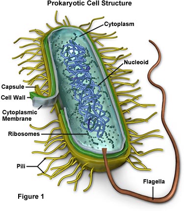
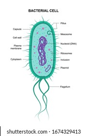
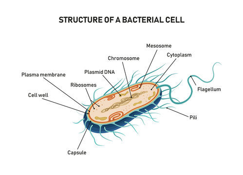
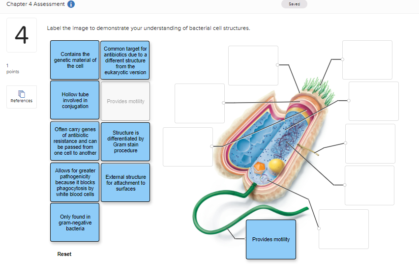
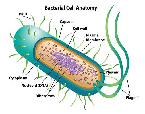
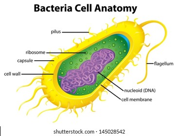
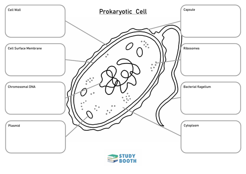
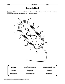
Bacterial cell picture with labels. Bacteria in Photos - photo gallery of bacteria facultatively anaerobic bacteria: Motility: nonmotile: Catalase test: catalase-positive: Oxidase test: negative* Spores: non-spore forming * Some species (non-human isolates) are positive: Streptococcus: ... (the cells stain a weak Gram-negative) Microscopic appearance: Spirochetes: Oxygen relationship: microaerobic: Motility: motile: Catalase ... Bacteria Cell Structures with labels - Dreamstime Get 15 images free trial Bacteria Cell Structures with labels Royalty-Free Vector Bacterial cell structures labeled on a bacillus cell with nucleoid DNA and ribosomes. External structures include the capsule, pili, and flagellum. Morphology of internal structures of bacteria. cell anatomy bacteria, prokaryotic cell, cell, internal structures, Label the Bacterium Cell - EnchantedLearning.com The cell is the basic unit of life. The following is a glossary of Bacterium cell terms. basal body - A structure that anchors the base of the flagellum and allows it to rotate. capsule - A layer on the outside of the cell wall. Most but not all bacteria have a capsule. cell wall - A thin membrane located outside the plasma membrane and within ... A Labeled Diagram of the Animal Cell and its Organelles A Labeled Diagram of the Animal Cell and its Organelles There are two types of cells - Prokaryotic and Eucaryotic. Eukaryotic cells are larger, more complex, and have evolved more recently than prokaryotes. Where, prokaryotes are just bacteria and archaea, eukaryotes are literally everything else.
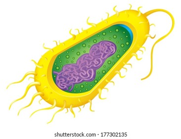
Structure of a bacterial cell, labeled. Stock Illustration Up to 500,000 print-runs. Not to be used on merchandise for sale. Extended license (US$79.99) Unlimited web, social, email, mobile views. More than 500,000 print-runs. Can be used on merchandise for sale. Download free with trial. Virus vs. Bacteria: What is the Difference? | Merriam-Webster Staphylococcus aureus bacterium labeled diagram. poster Staphylococcus aureus bacterium labeled diagram. poster Paper Type: Value Poster Paper (Matte) Your walls are a reflection of your personality, so let them speak with your favorite quotes, art, or designs printed on our custom Giclee posters! Choose from up to 5 unique, high quality paper types to meet your creative or business needs. DNA ligase - Wikipedia DNA ligase is a specific type of enzyme, a ligase, (EC 6.5.1.1) that facilitates the joining of DNA strands together by catalyzing the formation of a phosphodiester bond.It plays a role in repairing single-strand breaks in duplex DNA in living organisms, but some forms (such as DNA ligase IV) may specifically repair double-strand breaks (i.e. a break in both complementary strands of DNA).
600+ Free Bacteria & Virus Images - Pixabay 639 Free images of Bacteria Related Images:virusinfectionhealthcoronavirusdiseasemedicalbiologycovid-19medicine Bacteria and virus high resolution images. Find your perfect picture for your project. 361114 bacteriaillnessvirus 31566 koli bacteria 23073 bacteriamicrobiology 705182 virusmicroscope 30281 coronasprayearth 30570 monsterblueinternet Bacteria Labeled Illustrations & Vectors - Dreamstime Bacteria Labeled Stock Illustrations - 224 Bacteria Labeled Stock Illustrations, Vectors & Clipart - Dreamstime Bacteria Labeled Illustrations & Vectors Most relevant Best selling Latest uploads Within Results People Pricing License Media Properties More Safe Search 224 bacteria labeled illustrations & vectors are available royalty-free. Next page Intestinal transgene delivery with native E. coli ... - Cell Aug 04, 2022 · Live bacterial therapeutics (LBTs) could reverse diseases by engrafting in the gut and providing persistent beneficial functions in the host. However, attempts to functionally manipulate the gut microbiome of conventionally raised (CR) hosts have been unsuccessful because engineered microbial organisms (i.e., chassis) have difficulty in colonizing the hostile luminal environment. PHOTO GALLERY OF BACTERIA - Microbiology in pictures (the cells stain a weak Gram-negative) Microscopic appearance: Spirochetes: Oxygen relationship: microaerobic: Motility: motile: Catalase test:-Oxidase test:- ... Colonies of various bacteria. Bacteria photos. PICTURE OF THE MONTH. BACTERIA 2013 DECEMBER. DISK DIFFUSION METHOD FOR TESTING OF ANTIBIOTIC SUSCEPTIBILITY OF BACTERIA:
Anaerobic: MedlinePlus Medical Encyclopedia
Sequencing and Computational Approaches to Identification and ... May 20, 2013 · For example, retro-analysis of 202 published bacterial and viral metagenomes using a recently developed computational program DeconSeq, revealed the presence of human DNA contaminations in 64% of the metagenomes.21 This will definitely help in improving the accuracy of draft genome sequences. With improvements in draft genome sequences, better ...
2,325 Animal cell labeled Images, Stock Photos & Vectors - Shutterstock Animal cell labeled royalty-free images 2,325 animal cell labeled stock photos, vectors, and illustrations are available royalty-free. See animal cell labeled stock video clips Image type Orientation Color People Artists More Sort by Popular Biology Animals and Wildlife Healthcare and Medical Cooking Software eukaryote cell in vitro experiment
3 Common Bacteria Shapes - ThoughtCo Bacteria Shapes The three basic shapes of bacteria include cocci (blue), bacilli (green), and spirochetes (red). PASIEKA/Science Photo Library/Getty Images By Regina Bailey Updated on August 20, 2019 Bacteria are single-celled, prokaryotic organisms that come in different shapes.
Bacterial Cell Structure Labeling Diagram | Quizlet Bacterial Cell Structure Labeling 4.0 (1 review) + − Flashcards Learn Test Match Created by adkelly22 Terms in this set (16) Cytoplasm Water-based solution filling the entire cell Ribosomes Tiny particles composed of protein and RNA that are the sites of protein synthesis Nucleoid Composed of condensed DNA molecules. Cell Membrane
Animal Cell Labeled Diagram Pictures, Images and Stock Photos Browse 19 animal cell labeled diagram stock photos and images available, or start a new search to explore more stock photos and images. Newest results. Diagrams of animal and plant cells. Labelled diagrams of typical animal and plant cells with editable layers. Golgi apparatus or Golgi body.
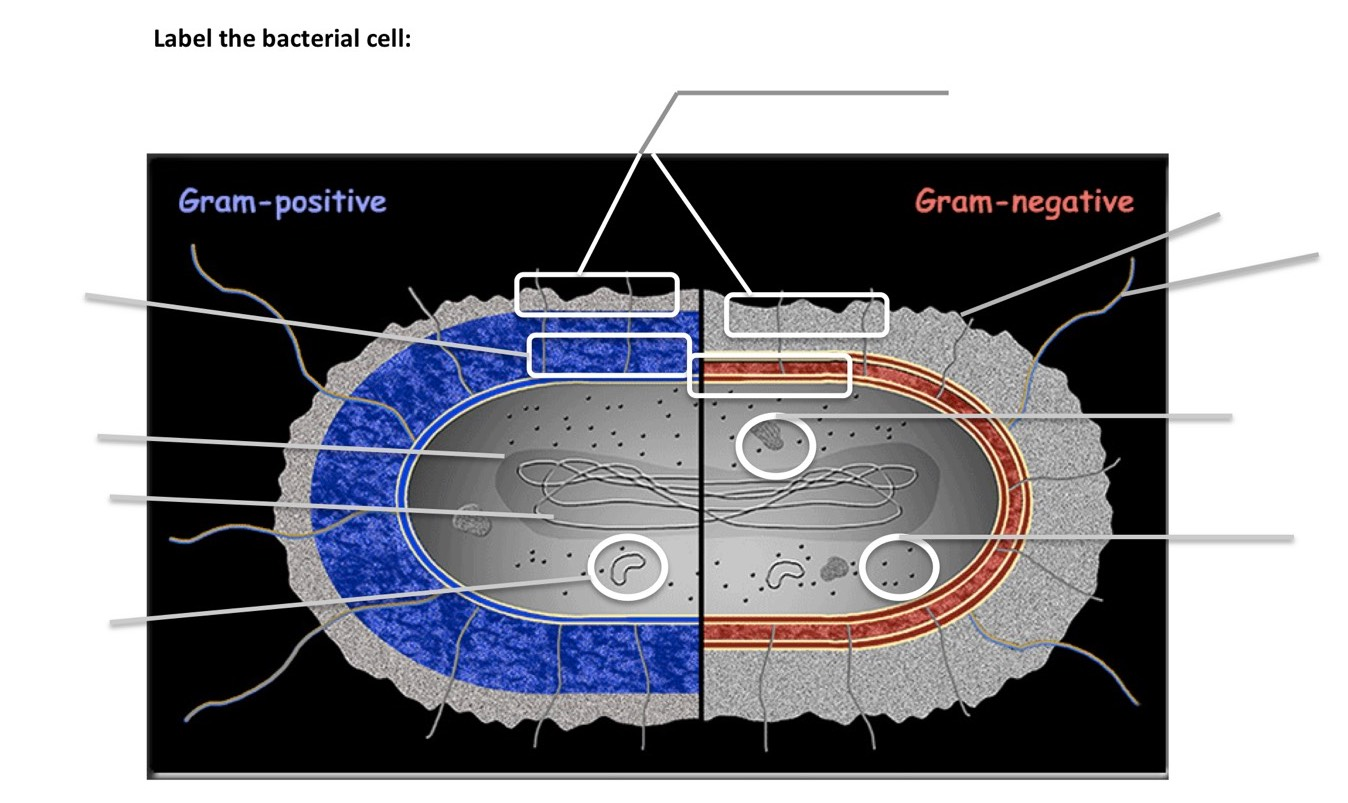
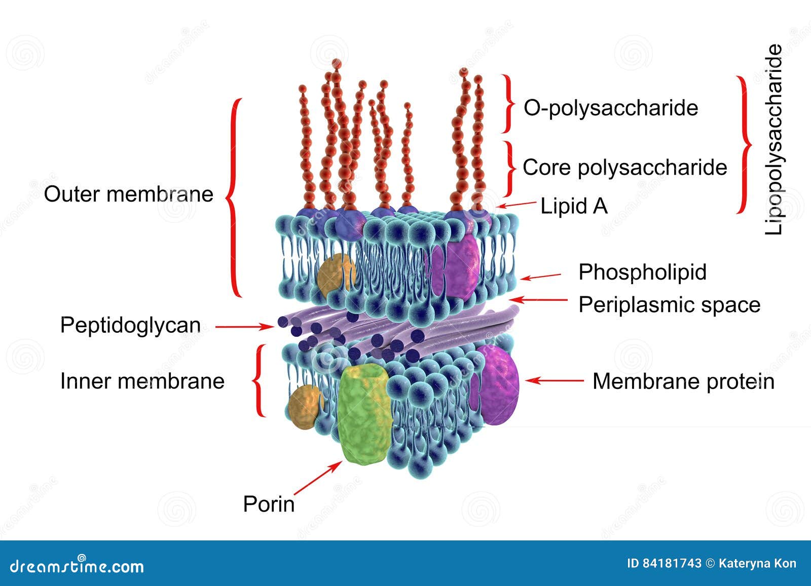
Structure of Bacteria (With Diagram) | Microbiology - Biology Discussion According to Peberdy (1980) the only compound present in the cell walls of both Gram-negative and Gram-positive bacteria is 'peptidoglycan'. The cell walls of Gram-positive bacteria contain up to 95% peptidoglycan and up to 10% teichoic acids. 9. Cytoplasmic membrane is a thin (5-10 nm) layer lining the inner surface of the cell wall.
Hyphae Overview, Function & Types | What is Hyphae in Fungi ... Jan 10, 2022 · Picture a fruit bowl, filled with ripe oranges and basking in the bright light of a sunny kitchen. ... This new cell will form a strand of hyphae, complete with a nucleus, organelles and cytoplasm ...
Image Library | CDC Online Newsroom | CDC Under a high magnification of 21674X, this digitally-colorized, scanning electron microscopic (SEM) image depicts a view of a dividing, Escherichia coli bacterium, clearly displaying the point at which the bacteria's cell wall was splitting into two separate organisms. See PHIL 7137 for a black and white version of this image. Listeria
Plant and Animal Cells - Labeled Graphics - The Biology Corner Plant Cell Lab Makeup - microscope observation of onion and elodea, if students missed the lab that day they can view a site with pictures to complete lab handout. Plant Cell Virtual Lab - use a virtual microscope to view plant cells. Comparing Plant and Animal Cells - looks at cheek and onion cells. Prokaryote Coloring - color a typical ...
Bacteria shapes and arrangements pictures Flashcards | Quizlet Start studying Bacteria shapes and arrangements pictures. Learn vocabulary, terms, and more with flashcards, games, and other study tools. ... If you had a bacteria infection would rather have a gram + bacteria or a gram -? ... Name the type of reproduction in which the offspring are identical to the parent cell. asexual reproduction. What ...
Animal Cell Labeled Pictures, Images and Stock Photos Browse 116 animal cell labeled stock photos and images available, or start a new search to explore more stock photos and images. Newest results Components of Eukaryotic cell, nucleus and organelles and plasma... Diagrams of animal and plant cells Labelled diagrams of typical animal and plant cells with editable layers. Golgi apparatus or Golgi body
PubMed PubMed® comprises more than 34 million citations for biomedical literature from MEDLINE, life science journals, and online books. Citations may include links to full text content from PubMed Central and publisher web sites.
Different Size, Shape and Arrangement of Bacterial Cells When viewed under light microscope, most bacteria appear in variations of three major shapes: the rod (bacillus), the sphere (coccus) and the spiral type (vibrio). In fact, structure of bacteria has two aspects, arrangement and shape. So far as the arrangement is concerned, it may Paired (diplo), Grape-like clusters (staphylo) or Chains (strepto).
Labeled Plant Cell With Diagrams | Science Trends Plant cells contain many organelles such as ribosomes, the nucleus, the plasma membrane, the cell wall, mitochondria, and chloroplasts. In addition, plant cells differ from animal cells in a number of key ways. Examining a diagram of the plant cell will help make the differences clearer. Let's go over the individual components of plant cells ...
Bacterial Cell Anatomy Labeling Structures On Stock ... - Shutterstock Stock Vector ID: 1523599223 Bacterial cell anatomy labeling structures on a bacillus cell with nucleoid DNA and ribosomes. External structures include the capsule, pili, and flagellum. Vector Formats EPS 2800 × 2193 pixels • 9.3 × 7.3 in • DPI 300 • JPG Show more Vector Contributor O OSweetNature Similar images See all
Bacteria - Definition, Structure, Diagram, Classification - BYJUS The bacteria diagram given below represents the structure of a typical bacterial cell with its different parts. The cell wall, plasmid, cytoplasm and flagella are clearly marked in the diagram. Bacteria Diagram representing the Structure of Bacteria Ultrastructure of a Bacteria Cell The structure of bacteria is known for its simple body design.
97,783 Bacteria Cell Stock Photos and Images - 123RF Bacteria Cell Stock Photos And Images 97,783 matches Page of 978 Structure of a bacterial cell. Anatomy of the prokaryote. unicellular organism. Vector diagram for your design, educational, medical, biological and science use Bacteria vector icon isolated on transparent background, Bacteria logo concept
Bacterial Staining Microbiology Images: Photographs and Videos of Gram ... 1. Endospore stain of Bacillus subtilis showing both endospores (green) & vegetative cells (pink) @1000xTM; 2. Negative endospore stain showing only vegetative cells @1000xTM; 3. Malachite green primary staining step of endopore stain with slide being heated over water bath; 4. Applying counterstain (safrinin) to bacterial smear as last step of ...
Structure of Bacterial Cell (With Diagram) - Biology Discussion It is a tough and rigid structure of peptidoglycan with accessory specific materials (e.g. LPS, teichoic acid etc.) surrounding the bacterium like a shell and lies external to the cytoplasmic membrane. It is 10-25 nm in thickness. It gives shape to the cell. Nucleus: The single circular double-stranded chromosome is the bacterial genome.
Eubacteria, True bacteria, Bacteria -- Discover Life mobile
Bacterial cells - Cell structure - Edexcel - GCSE Combined Science ... Feature Eukaryotic cell (plant and animal cell) Prokaryotic cell (bacterial cell) Size: Most are 5 μm - 100 μm: Most are 0.2 μm - 2.0 μm: Outer layers of cell

/shigella-bacteria--illustration-758308491-5a02252f9e9427003c1759be.jpg)














Post a Comment for "41 bacterial cell picture with labels"