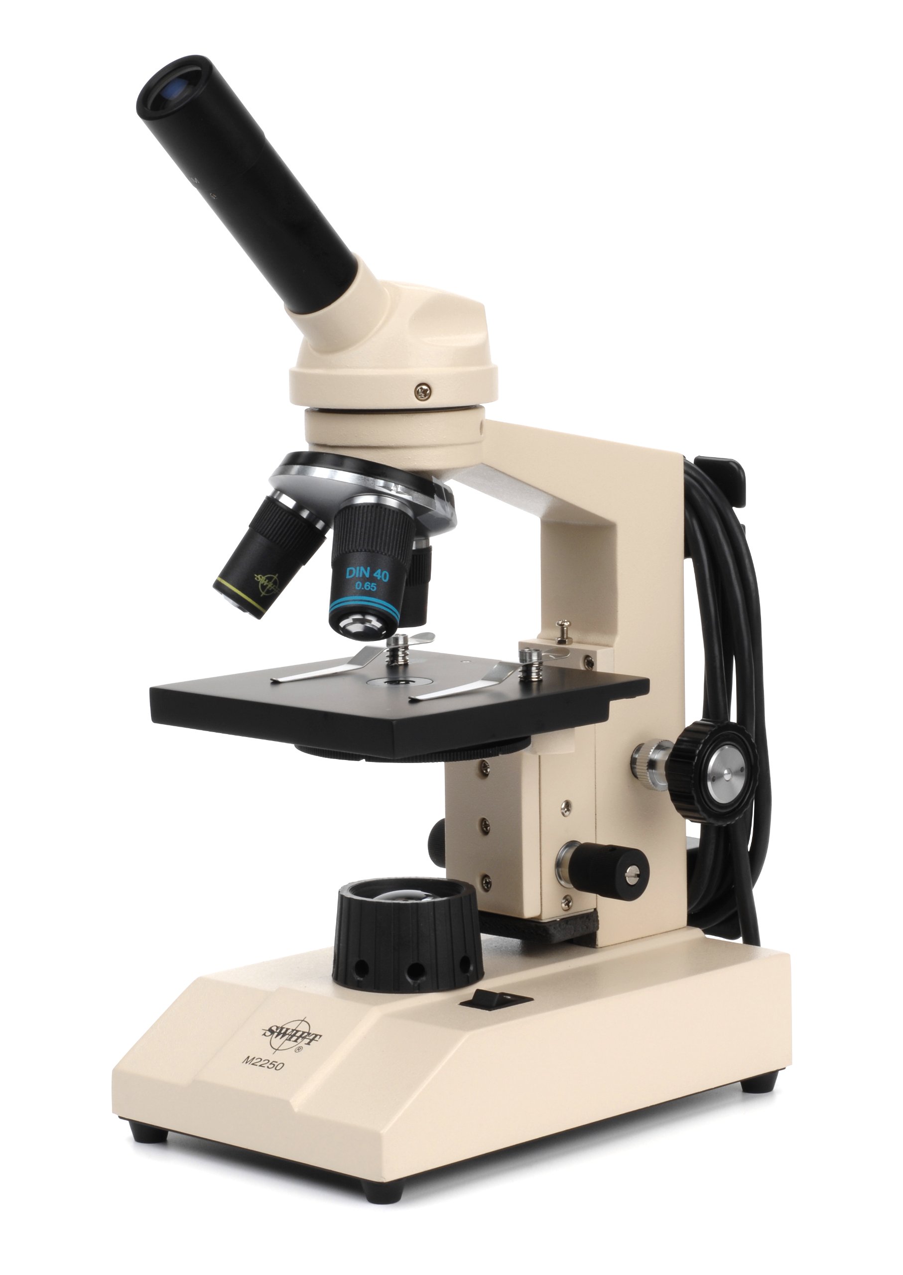
42 light microscope with labels
Microscope Labeling Game - PurposeGames.com An unregistered player played the game 1 hour ago About this Quiz This is an online quiz called Microscope Labeling Game There is a printable worksheet available for download here so you can take the quiz with pen and paper. This quiz has tags. Click on the tags below to find other quizzes on the same subject. Science microsope Your Skills & Rank Microscope Types (with labeled diagrams) and Functions This is an advanced microscope that has specific application in viewing, observing and measuring the optical thickness and phase of completely transparent specimens and objects. A tiny interferometer is used and a specimen is placed on beam path of it. This path is split and then rejoined to create two superimposed images of the specimen in focus.
Label a Microscope - Storyboard That Knowing the names of the different parts of the microscope is essential to be able to use one properly. Create a poster that labels the parts of a microscope and includes descriptions of what each part does. Click "Start Assignment". Use a landscape poster layout (large or small). Search for a diagram of a microscope.

Light microscope with labels
Microscope, Microscope Parts, Labeled Diagram, and Functions Majority of high quality microscopes used in laboratory include an Abbe condenser with an iris diaphragm. When iris diaphragm is combined with Abbe condenser, it control both the quantity of light applied as well as focus on the specimen. Aperture: It is the hole in the stage through which the base (transmitted) light reaches the stage. Compound Light Microscope Labeling - Printable About this Worksheet. This is a free printable worksheet in PDF format and holds a printable version of the quiz Compound Light Microscope Labeling. By printing out this quiz and taking it with pen and paper creates for a good variation to only playing it online. Labelled Diagram Of A Light Microscope | Products & Suppliers ... Products/Services for Labelled Diagram Of A Light Microscope Microscopes - (705 companies) ...and transmission electron microscopes. Acoustic and ultrasonic microscopes use sound waves to create images of the sample. Compound microscopes use a single light path. These types of microscopes can have a single eyepiece (monocular) or a dual eyepiece...
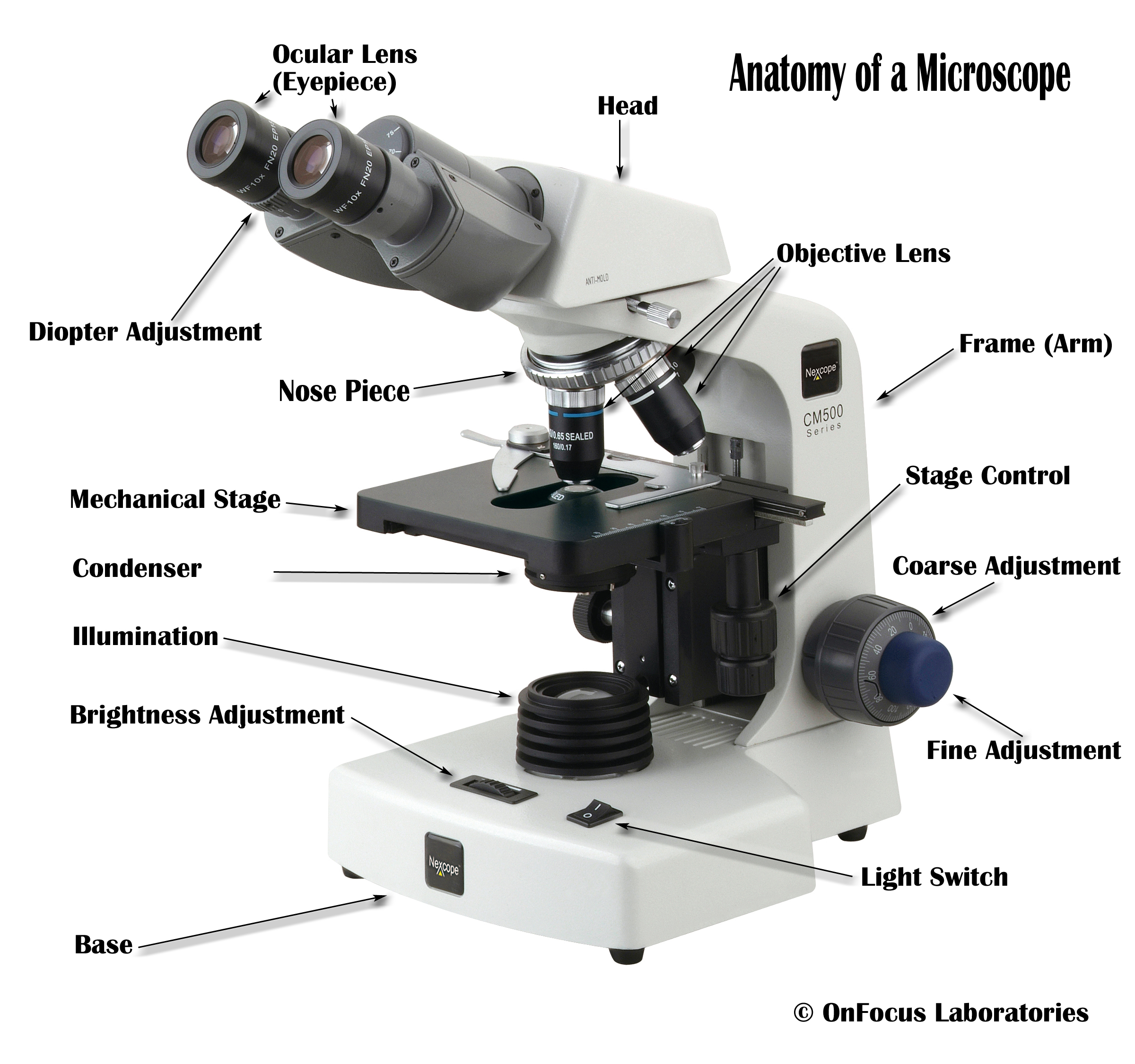
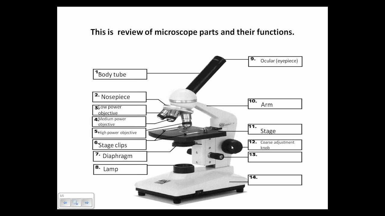
Light microscope with labels. Compound Microscope Parts, Functions, and Labeled Diagram Compound Microscope Definitions for Labels. Eyepiece (ocular lens) with or without Pointer: The part that is looked through at the top of the compound microscope. Eyepieces typically have a magnification between 5x & 30x. Monocular or Binocular Head: Structural support that holds & connects the eyepieces to the objective lenses. Compound Microscope: Definition, Diagram, Parts, Uses, Working ... - BYJUS A compound microscope is defined as. A microscope with a high resolution and uses two sets of lenses providing a 2-dimensional image of the sample. The term compound refers to the usage of more than one lens in the microscope. Also, the compound microscope is one of the types of optical microscopes. The other type of optical microscope is a ... Parts of the Microscope with Labeling (also Free Printouts) Parts of the Microscope with Labeling (also Free Printouts) A microscope is one of the invaluable tools in the laboratory setting. It is used to observe things that cannot be seen by the naked eye. Table of Contents 1. Eyepiece 2. Body tube/Head 3. Turret/Nose piece 4. Objective lenses 5. Knobs (fine and coarse) 6. Stage and stage clips 7. Aperture Microscope Labeling - The Biology Corner 1) Start with scanning (the shortest objective) and only use the COARSE knob . Once it is focused… 2) Switch to low power (medium) and only use the COARSE knob . You may need to recenter your slide. Once it is focused.. 3) Switch to high power (long objective).
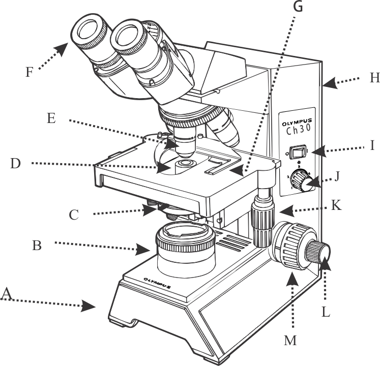
Explanation and Labelled Images - New York Microscope Company A fluorescence microscope is used to study organic and inorganic samples. Fluorescence microscopy uses fluorescence and phosphorescence to examine the structural organization, spatial distribution of samples. It is particularly used to study samples that are complex and cannot be examined under conventional transmitted-light microscope. Parts of a microscope with functions and labeled diagram Apr 19, 2022 · Head – This is also known as the body. It carries the optical parts in the upper part of the microscope. Base – It acts as microscopes support. It also carries microscopic illuminators. Arms – This is the part connecting the base and to the head and the eyepiece tube to the base of the microscope. Label the Light Microscope - Labelled diagram - Wordwall Drag and drop the pins to their correct place on the image.. Eyepiece, Light Source, Base, Stage, Stage Clips, Fine Focus, Coarse Focus, Arm, Objective Lens. Microscope | Biology I Laboratory Manual | | Course Hero View the slide with your eyes, and then place it onto the microscope. Use the focusing sequence to view the slide under low power. Procedure Draw the letter "e" as it appears when you look at the slide without the microscope. Draw the letter "e" as it appears when you look at the slide under the microscope.
Light Microscope- Definition, Principle, Types, Parts ... Apr 07, 2022 · A light microscope is a biology laboratory instrument or tool, that uses visible light to detect and magnify very small objects and enlarge them. They use lenses to focus light on the specimen, magnifying it thus producing an image. The specimen is normally placed close to the microscopic lens. Light Microscope-Definition, Principle, Types, Parts, Labeled ... Jun 29, 2022 · A light microscope is a device or instrument used in biology laboratories that uses visible light to locate, magnify, and expand micro objects. Using lenses, they focus light on the specimen and magnify it to generate a photograph. Typically, the specimen is positioned close to the microscopic lens. The kinds and quantity of lenses that make up ... Microscope labels Flashcards | Quizlet Laurxyala Microscope label Terms in this set (14) ocular lens / eyepiece diopter adjustment Arm Coarse focus Fine focus On/off switch Base light source iris diaphragm Condenser Stage slide holder Objective lens Nose piece 10 terms Centrifuge Skills Test 14 terms Laurxyala Abbreviation 1 VTA 170 instruments Laurxyala Microscope Parts, Function, & Labeled Diagram - slidingmotion Condenser. The condenser is to focus the light, which passes from the microscopic illuminator to the specimen. This condenser is located just below the diaphragm. This diaphragm is one of the important parts of the compound microscope which will help to get an accurate and sharp image. The condenser has a magnification power of 400X and above.
Compound Light Microscope Labeled - Made By Creative Label
Light microscopes - Cell structure - Edexcel - BBC Bitesize Microscopes are used to produce magnified images. There are two main types of microscope: light microscopes are used to study living cells and for regular use when relatively low magnification and...
Labeling A Compound Light Microscope - ClipArt Best
A Study of the Microscope and its Functions With a Labeled Diagram The microscope is an important instrument in the world of biological science. Diagrams have always been of great help in understanding both the structural and functional aspects of entities. These labeled microscope diagrams and the functions of its various parts, attempt to simplify the microscope for you.
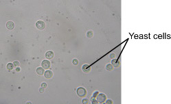
Yeast Bubbles - Experiments on Microscopes 4 Schools
Labeling the Parts of the Microscope Labeling the Parts of the Microscope This activity has been designed for use in homes and schools. Each microscope layout (both blank and the version with answers) are available as PDF downloads. You can view a more in-depth review of each part of the microscope here. Download the Label the Parts of the Microscope PDF printable version here.

Review Questions
Parts of Stereo Microscope (Dissecting microscope) - labeled diagram ... Labeled part diagram of a stereo microscope Major structural parts of a stereo microscope. There are three major structural parts of a stereo microscope. The viewing Head includes the upper part of the microscope, which houses the most critical optical components, including the eyepiece, objective lens, and light source of the microscope.

Light
Compound Microscope Parts - Labeled Diagram and their Functions The eyepiece (or ocular lens) is the lens part at the top of a microscope that the viewer looks through. The standard eyepiece has a magnification of 10x. You may exchange with an optional eyepiece ranging from 5x - 30x. [In this figure] The structure inside an eyepiece. The current design of the eyepiece is no longer a single convex lens.

Simple Light Microscope Labeled - Micropedia
Light Microscope Labeled - how scanning electron microscopes work ... Light Microscope Labeled - 16 images - senior biology cell theory microscopy, what is a light microscope with pictures, 29 you will love labeling a compound microscope db, microscope imaging station gallery,
Label a microscope - Teaching resources
Compound Light Microscope: Everything You Need to Know A fluorescence microscope, also called a confocal microscope, is a kind of biological microscope that operates by using different light colors and wavelengths over-dyed specimen samples in order for the dye to interact with the light, after which the resulting image is scanned.
1.Fully label the compound microscope below.
Label the microscope — Science Learning Hub All microscopes share features in common. In this interactive, you can label the different parts of a microscope. Use this with the Microscope parts activity to help students identify and label the main parts of a microscope and then describe their functions. Drag and drop the text labels onto the microscope diagram.
Labeling A Compound Light Microscope - ClipArt Best
Student's Guide: How to Use a Light Microscope An ultraviolet microscope uses UV light to view specimens at a resolution that isn't possible with the common brightfield microscope. It utilizes UV optics, light sources, as well as cameras. Because of the shorter wavelengths of UV light (180-400 nm), the image produced is clearer and more distinct at a magnification approximately double what ...
Microscope Diagram to Print
Microscope Parts and Functions With Labeled Diagram and ... First, the purpose of a microscope is to magnify a small object or to magnify the fine details of a larger object in order to examine minute specimens that cannot be seen by the naked eye. Here are the important compound microscope parts... Eyepiece: The lens the viewer looks through to see the specimen.
Labeled Compound Light Microscope - ClipArt Best
Simple Microscope - Diagram (Parts labelled), Principle, Formula and Uses A simple microscope consists of Optical parts Mechanical parts Labeled Diagram of simple microscope parts Optical parts The optical parts of a simple microscope include Lens Mirror Eyepiece Lens A simple microscope uses biconvex lens to magnify the image of a specimen under focus.

Search in gallery
Sperm Under Microscope with Labeled Diagram Under the light microscope, the sperm consists of two main portions - the head and the tail. But, the electron microscope shows four different parts in the tail of spermatozoa. ... So, this article provides the details structural features of sperm under the light microscope. All the labeled diagrams might help you identify the sperms from ...

Cells
Microscope Labeling - The Biology Corner Students label the parts of the microscope in this photo of a basic laboratory light microscope. Can be used for practice or as a quiz. Name_____ Microscope Labeling . Microscope Use: 15. When focusing a specimen, you should always start with the _____ objective.
Microscope Review.wmv - YouTube
Labelled Diagram Of A Light Microscope | Products & Suppliers ... Products/Services for Labelled Diagram Of A Light Microscope Microscopes - (705 companies) ...and transmission electron microscopes. Acoustic and ultrasonic microscopes use sound waves to create images of the sample. Compound microscopes use a single light path. These types of microscopes can have a single eyepiece (monocular) or a dual eyepiece...
Microscope Clip Art at Clker.com - vector clip art online, royalty free & public domain
Compound Light Microscope Labeling - Printable About this Worksheet. This is a free printable worksheet in PDF format and holds a printable version of the quiz Compound Light Microscope Labeling. By printing out this quiz and taking it with pen and paper creates for a good variation to only playing it online.

Anatomy and Physiology I Coursework: Microscope A+P
Microscope, Microscope Parts, Labeled Diagram, and Functions Majority of high quality microscopes used in laboratory include an Abbe condenser with an iris diaphragm. When iris diaphragm is combined with Abbe condenser, it control both the quantity of light applied as well as focus on the specimen. Aperture: It is the hole in the stage through which the base (transmitted) light reaches the stage.
Search in gallery

Microscope World Blog: Carpet under the Microscope
Post a Comment for "42 light microscope with labels"