42 brain pictures and labels
Brain Label (Remote) - The Biology Corner This brain labeling activity was created for remote learners as an alternative to the labeling and coloring worksheet we would traditionally do in class. Instead of coloring and labeling on printouts, students use google slides to drag labels to the images or type the answers into text boxes. Whole Brain Segmentation and Labeling from CT Using Synthetic MR Images ... With MALP-EM processing of the ground truth T1-w as the reference, we compute the Dice coefficient between multi-atlas segmentations using either the subject CT images with MV label fusion (red), or synthetic T1-w with MV (green) or JLF (blue), as the label fusion, and MALP-EM (yellow).Note that MALP-EM uses the OASIS atlas with manually delineated labels, while the other three use the ...
How to Draw a Brain: 14 Steps (with Pictures) - wikiHow Once you've drawn the cartoon or realistic brain, you can go back and add color or label the parts. Method 1 Sketching an Easy Cartoon Brain 1 Draw a large bean shape to make the outline for the brain. Use a pencil to sketch a kidney bean shape on your paper. You can make the outline for the brain any size you like.

Brain pictures and labels
Picture of the Brain - WebMD • The cerebellum is at the base and the back of the brain. The cerebellum is responsible for coordination and balance. The brain is also divided into several lobes: • The frontal lobes are... Labeling Brain Structures - John Muschelli 1 Labels in template space. In Processing Within-Visit MRI, we registered the T1 image to the Eve template using a non-linear registration (SyN) (Avants et al. 2008). Also, we applied this transformation to the intensity-normalized T1, T2, and FLAIR images, so that these image are located in the same space as the Eve atlases. We can overlay the ... 3D Brain This interactive brain model is powered by the Wellcome Trustand developed by Matt Wimsattand Jack Simpson; reviewed by John Morrison, Patrick Hof, and Edward Lein. Structure descriptions were written by Levi Gadye and Alexis Wnukand Jane Roskams. Copyright © Society for Neuroscience (2017). Users may copy images and text, but must provide ...
Brain pictures and labels. Human Brain Diagram Photos and Premium High Res Pictures - Getty Images 1,018 Human Brain Diagram Premium High Res Photos Browse 1,018 human brain diagram stock photos and images available, or start a new search to explore more stock photos and images. of 17 NEXT Labels of the brain, midbrain, pons, medulla, cervical spinal cord ... Labels of the brain, midbrain, pons, medulla, cervical spinal cord, thoracic spinal cord and skeletal muscle. Stock Illustration - LifeART. sa302024 Fotosearch Stock Photography and Stock Footage helps you find the perfect photo or footage, fast! We feature 71,900,000 royalty free photos, stock footage clips, digital videos, vector clip art images, clipart pictures, background graphics ... 1,000+ of the Best Brain Pictures for Free [HD] - Pixabay Pick the perfect brain picture for your project. HD to 4K quality, available for free on all devices! 1658 288. brain mind psychology. 1187 271. artificial intelligence. 704 133. artificial intelligence. 762 180. Whole Brain Segmentation and Labeling from CT Using Synthetic MR Images This paper presents a whole brain segmentation and labeling method for non-contrast CT images that first uses a fully convolutional network (FCN) to synthesize an MR image from a CT image and then ...
101 labeled brain images and a consistent human cortical labeling ... 101 labeled brain images and a consistent human cortical labeling protocol Abstract We introduce the Mindboggle-101 dataset, the largest and most complete set of free, publicly accessible, manually labeled human brain images. Nervous System - Label the Brain Nervous System - Label the Brain Nervous System - Brain Name: Choose the correct names for the parts of the brain. ( 1) (2) (3) (4) (5) (6) (7) (8) ( 9) This brain part controls thinking. (10) This brain part controls balance, movement, and coordination. (11) This brain part controls involuntary actions such as breathing, heartbeats, and digestion. 75,682 Brain Anatomy Stock Photos and Images - 123RF Brain Anatomy Stock Photos And Images 75,682 matches Page of 757 Brain lobes vector illustration. Human brain infographic vector. Brain lobes functions Serotonin pathway. Humans brain with serotonin pathways. psychiatric and neurological disorders. 3D render of a medical image showing male figure with brain tumour Neocortex vector illustration. Diagram of the Brain and its Functions - Bodytomy Functions. The frontal lobe is involved with the main executive functions of the brain, which include: Judgment, that is, the ability to recognize future consequences resulting from ongoing actions. This activity mostly occurs in the pre-frontal area. Analytical and critical reasoning.
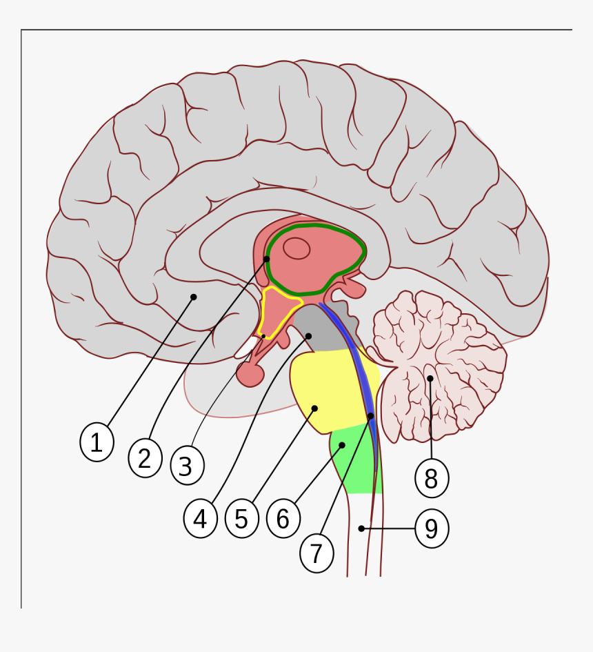
101 Labeled Brain Images and a Consistent Human Cortical Labeling ... In this article we introduce this dataset of manually edited brain image labels applied to the T1-weighted MR images of publicly available multi-modal data acquired from healthy individuals. We also introduce a benchmark for the evaluation of automated registration/segmentation/labeling methods by comparing the manual labels according to this "Desikan-Killiany-Tourville" (DKT) protocol with automatically generated labels. Parts of the brain: Learn with diagrams and quizzes - Kenhub Labeled brain diagram First up, have a look at the labeled brain structures on the image below. Try to memorize the name and location of each structure, then proceed to test yourself with the blank brain diagram provided below. Labeled diagram showing the main parts of the brain Blank brain diagram (free download!) The Brain - Diagram and Explanation AMYGDALA: Lying deep in the center of the limbic emotional brain, this powerful structure, the size and shape of an almond, is constantly alert to the needs of basic survival including sex, emotional reactions such as anger and fear. Consequently it inspires aversive cues, such as sweaty palms, and has recently been associated with a range of mental conditions including depression to even autism. Human Brain Photos and Premium High Res Pictures - Getty Images human brain sensory 27,942 Human Brain Premium High Res Photos Browse 27,942 human brain stock photos and images available, or search for human brain anatomy or human brain illustration to find more great stock photos and pictures. Related searches: human brain anatomy human brain illustration human brain diagram human brain scan human brain vector
Brain Label | Human anatomy and physiology, Basic anatomy and ... Brain Label Image of the brain showing its major features for students to practice labeling. Answers are included. Biologycorner 17k followers More information Brain Label Find this Pin and more on Anatomy & Physiology by Page Johnson. Basic Anatomy And Physiology Brain Anatomy Science Education Physical Science Science Experiments
Brain: Atlas of human anatomy with MRI - e-Anatomy - IMAIOS MRI Atlas of the Brain. This page presents a comprehensive series of labeled axial, sagittal and coronal images from a normal human brain magnetic resonance imaging exam. This MRI brain cross-sectional anatomy tool serves as a reference atlas to guide radiologists and researchers in the accurate identification of the brain structures.
Label Brain Diagram Printout - EnchantedLearning.com Read the definitions below, then label the brain anatomy diagram. Cerebellum - the part of the brain below the back of the cerebrum. It regulates balance, posture, movement, and muscle coordination. Corpus Callosum - a large bundle of nerve fibers that connect the left and right cerebral hemispheres.
2,782 Labeled brain anatomy Images, Stock Photos & Vectors - Shutterstock Find Labeled brain anatomy stock images in HD and millions of other royalty-free stock photos, illustrations and vectors in the Shutterstock collection. Thousands of new, high-quality pictures added every day.
Parts of the Brain Activity for Kids, Brain Diagram, and Worksheets for ... PARIETAL LOBES - The parietal lobe provides sensory information to the brain including touch, pain and temperature. OCCIPITAL LOBES - The occipital lobe processes and interprets everything we see TEMPORAL LOBES - The temporal lobe controls emotions and short-term memory
Neuroscience Resources for Kids - Coloring Book Color these pictures and make your own coloring book filled with drawings about neuroscience. ... Then click on the "print" button of your browser to print out the picture. Color the picture and label the drawing if you like. ... Spinal Cord: Neuron: Neuron: Brain. Brain (Top View) Brain (Bottom View) Brain (Side View) Bipolar Neuron: Unipolar ...
Human Brain Stock Photos, Pictures & Royalty-Free Images - iStock Browse 230,634 human brain stock photos and images available, or search for human brain anatomy or human brain illustration to find more great stock photos and pictures. Human brain icon in line style. Human brain icon in line style. For your design, logo. Vector illustration. Editable Stroke. AI (Artificial Intelligence) concept. Deep learning.
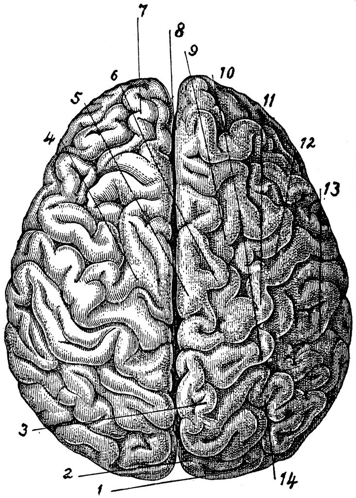
Illustration Picture of Brain Anatomy - Brain - eMedicineHealth Medical Illustrations Picture of Brain The brain is the complex organ responsible for processing sensory information (sound, touch, taste, sight, and smell). The brain controls voluntary and involuntary movements. Signals from the brain tell muscles to contract. Input from the brain controls the function of other organs in the body.
Human Brain Worksheets - Superstar Worksheets Learning about the human brain is a fascinating area of study for students of all ages. Use these free printable human brain worksheets for your science notebooks, journals, and science projects. ... This is a simple worksheet for children to color and label the different areas of the human brain. ... Mystery Picture Pixel Coloring Pages ...
3D Brain This interactive brain model is powered by the Wellcome Trustand developed by Matt Wimsattand Jack Simpson; reviewed by John Morrison, Patrick Hof, and Edward Lein. Structure descriptions were written by Levi Gadye and Alexis Wnukand Jane Roskams. Copyright © Society for Neuroscience (2017). Users may copy images and text, but must provide ...
Labeling Brain Structures - John Muschelli 1 Labels in template space. In Processing Within-Visit MRI, we registered the T1 image to the Eve template using a non-linear registration (SyN) (Avants et al. 2008). Also, we applied this transformation to the intensity-normalized T1, T2, and FLAIR images, so that these image are located in the same space as the Eve atlases. We can overlay the ...
Picture of the Brain - WebMD • The cerebellum is at the base and the back of the brain. The cerebellum is responsible for coordination and balance. The brain is also divided into several lobes: • The frontal lobes are...


Post a Comment for "42 brain pictures and labels"