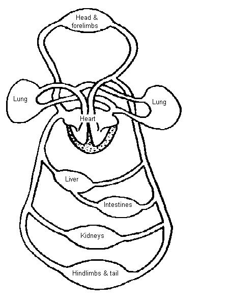
45 heart structure with labels
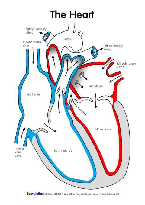
Structure of the Heart | SEER Training Layers of the Heart Wall Three layers of tissue form the heart wall. The outer layer of the heart wall is the epicardium, the middle layer is the myocardium, and the inner layer is the endocardium. Chambers of the Heart The internal cavity of the heart is divided into four chambers: Right atrium Right ventricle Left atrium Left ventricle Label Heart Anatomy Diagram Printout - EnchantedLearning.com Every day, the heart pumps about 2,000 gallons (7,600 liters) of blood, beating about 100,000 times. Label the heart anatomy diagram below using the heart glossary. Note: On the diagram, the right side of the heart appears on the left side of the picture (and vice versa) because you are looking at the heart from the front.
Label the heart - Science Learning Hub Label the heart — Science Learning Hub Label the heart Add to collection In this interactive, you can label parts of the human heart. Drag and drop the text labels onto the boxes next to the diagram. Selecting or hovering over a box will highlight each area in the diagram. Pulmonary vein Right atrium Semilunar valve Left ventricle Vena cava

Heart structure with labels
The Heart and Circulation of Blood - LSA The center of the circulatory system is the heart, which is the main pumping mechanism. The heart is made of muscle. The heart is shaped something like a cone, with a pointed bottom and a round top. It is hollow so that it can fill up with blood. An adult’s heart is about the size of a large orange and weighs a little less than a pound. The Anatomy of the Heart, Its Structures, and Functions The heart is the organ that helps supply blood and oxygen to all parts of the body. It is divided by a partition (or septum) into two halves. The halves are, in turn, divided into four chambers. The heart is situated within the chest cavity and surrounded by a fluid-filled sac called the pericardium. This amazing muscle produces electrical ... Human Heart - Anatomy, Functions and Facts about Heart The external structure of the heart has many blood vessels that form a network, with other major vessels emerging from within the structure. The blood vessels typically comprise the following: Veins supply deoxygenated blood to the heart via inferior and superior vena cava, and it eventually drains into the right atrium.
Heart structure with labels. Carbohydrates | American Heart Association Carbohydrates are either called simple or complex, depending on the food’s chemical structure and how quickly the sugar is digested and absorbed. The type of carbohydrates that you eat makes a difference – Foods that contain high amounts of simple sugars, especially fructose raise triglyceride levels. Heart Structure: Interactive Labelling Activity | Teaching ... File previews. ppt, 2.23 MB. An interactive PowerPoint on heart structure. Student prompted to label different part of the heart and includes fun noises! Tes classic free licence. BYJU'S Online learning Programs For K3, K10, K12, NEET ... BYJU'S Online learning Programs For K3, K10, K12, NEET ... PDF Anatomy of Heart Labeled and Unlabeled Images (a) Anterior view of the external heart C' 2019 Pearson Education. Aort'c arch Ligamentum arteriosum Left pulmonary artery Left pulmonary ve ns Auricle of left atrium Circumflex artery Left coronary artery (in atrioventricular sulcus) Great cardiac vein Left ventricle Anterior interventricular artery (in anterior interventricular sulcus) Apex
Structure of the Heart | Biology for Majors II The heart is composed of three layers; the epicardium, the myocardium, and the endocardium, illustrated in Figure 1. The inner wall of the heart has a lining called the endocardium.The myocardium consists of the heart muscle cells that make up the middle layer and the bulk of the heart wall. The outer layer of cells is called the epicardium, of which the second layer is a membranous layered ... Heart Diagram with Labels and Detailed Explanation The heart is located under the ribcage, between the lungs and above the diaphragm. It weighs about 10.5 ounces and is cone shaped in structure. It consists of the following parts: Heart Detailed Diagram Heart - Chambers There are four chambers of the heart . The upper two chambers are the auricles and the lower two are called ventricles. Easy way to draw heart structure by 5 steps | labeling of ... My youtube channel : facebook page : way to draw hea... Human Heart (Anatomy): Diagram, Function, Chambers ... The heart is a muscular organ about the size of a fist, located just behind and slightly left of the breastbone. The heart pumps blood through the network of arteries and veins called the...
PDF Heart Structure - in.gov The heart is an organ about the size of a fist. It is made of muscle and pumps blood through the body. Tube-like structures called blood vessels carry blood through the body and heart. The heart and blood vessels make up the cardiovascular system. Structure of the Heart The heart has four chambers: two upper chambers call Heart Anatomy: Labeled Diagram, Structures, Blood Flow ... There are 4 chambers, labeled 1-4 on the diagram below. To help simplify things, we can convert the heart into a square. We will then divide that square into 4 different boxes which will represent the 4 chambers of the heart. The boxes are numbered to correlate with the labeled chambers on the cartoon diagram. heart | Structure, Function, Diagram, Anatomy, & Facts ... heart, organ that serves as a pump to circulate the blood. It may be a straight tube, as in spiders and annelid worms, or a somewhat more elaborate structure with one or more receiving chambers (atria) and a main pumping chamber (ventricle), as in mollusks. In fishes the heart is a folded tube, with three or four enlarged areas that correspond to the chambers in the mammalian heart. Structure of the Heart - The Franklin Institute Structure of the Heart Although most people know that the human heart doesn't bear much resemblance to a heart drawn on a Valentine's Day card, the image can still be a useful way to learn and remember the parts of the heart.
Circulatory System Worksheet - WikiEducator
Label the Heart Diagram - Quizlet Label the Heart STUDY Learn Write Test PLAY Match Created by bluesas9 Terms in this set (15) Superior Vena Cava ... Right Ventricle ... Left Atrium ... Atrioventricular/Tricuspid Valve ... Atrioventricular/Mitral Valve ... Septum ... Right Atrium ... Semi-lunar Valves ... Left Pulmonary Veins ... Right Pulmonary Veins ... Left Pulmonary Arteries
Label the Heart Worksheets (SB6634) - SparkleBox
heart labeling Diagram - Quizlet A vein that is the second largest vein in the human body and returns blood to the right atrium of the heart from the upper half of the body. ... at the opening of the right atrium of the heart into the right ventricle and that resembles the mitral valve in structure but consists of three triangular membranous flaps.

Heart Disease: Structure Of The Heart
How to Draw the Internal Structure of the Heart (with ... To draw the internal structure of a human heart, follow the steps below. Part 1 Finding a Diagram 1 To find a good diagram, go to Google Images, and type in "The Internal Structure of the Human Heart". Find an image that displays the entire heart, and click on it to enlarge it. 2 Find a piece of paper and something to draw with.

Heart Structures Flashcards | Easy Notecards
Heart Information Center: Heart Anatomy | Texas Heart ... Your heart is located between your lungs in the middle of your chest, behind and slightly to the left of your breastbone. A double-layered membrane called the pericardium surrounds your heart like a sac. The heart weighs between 7 and 15 ounces (200 to 425 grams) and is a little larger than the size of your fist.
Regions Of Stomach Diagram , Free Transparent Clipart - ClipartKey
Human Heart - Anatomy, Functions and Facts about Heart The external structure of the heart has many blood vessels that form a network, with other major vessels emerging from within the structure. The blood vessels typically comprise the following: Veins supply deoxygenated blood to the heart via inferior and superior vena cava, and it eventually drains into the right atrium.
Post a Comment for "45 heart structure with labels"